

We all know what an X-Ray is, but do we really? We know that X-Rays are used in the dental and medical fields to help doctors and dentists see any problems that wouldn’t be noticed by the naked eye, but what’s really going on when we turn on that machine? Is it necessary? And most importantly just how dangerous are X-Rays?
Believe it or not, X-Rays (also known as “dental radiographs”) are a dentist’s most powerful tool when it comes to diagnosing and preventing potential risks and injuries. Dentists can clearly see things like cavities and cracked teeth and can quickly and efficiently address these issues.
What Is an X-Ray?
A common misconception of many people is that x-rays are a relatively newer discovery. But x-rays were actually first discovered in 1895 when the German scientist Wilhelm Conrad Roentgen performed various experiments on electrons. The name “x-ray” came from the ambiguous nature of them at the time. Nobody really knew what was going on or exactly how they worked, so the term “X” was used.
The very first successful X-Ray was made just a mere 14 days after the initial discovery. It wasn’t long before the magnitude of this new technology was realized and over the next century or so, professionals all over the globe have worked to develop the powerful, accurate, and safe x-rays we use today.
We know how they came to be, we know that they’re effective, but how do they work exactly? Well essentially an X-Ray is a light wave much like the light waves we see all around us on a regular basis. The difference between visible light waves and x-ray light waves is simply just the wavelength. In the same way we can only hear certain pitches of sound based on their wavelength, we can only see certain types of light based on their wavelength. Microwaves and radio waves are both additional examples of invisible light waves we interact with on a daily basis.
An x-ray machine basically just produces a concentrated beam of electrons known as x-ray photons. Because of the nature of x-rays, they have the unique capabilities to pass through non-metallic objects such as human tissue and organs.
In simpler terms, an x-ray machine is basically a over-sized camera that manipulates x-ray waves in a way that allows dentists to take pictures of your internal structures. This way dentists and doctors don’t have to cut you open to get their eyes on things and that is something I’m sure we are all thankful for.
Types of X-Rays
Essentially most types of x-rays in the dental field will fall under one of two categories: intraoral or extraoral. Intraoral meaning the x-ray is taken from inside of the mouth and extraoral meaning the x-ray is taken from the outside. But within those two categories there are several more specific types of x-rays that are commonly used in the dental practice. Below we’ve listed a few.
- Bitewing X-Rays: This type of x-ray is generally the most common in a dentist office and you’ve probably done quite a few of these in your lifetime. The name bitewing comes from the fact that you bite down on piece of material that acts as a sensor for the x-ray. This form of x-ray shows only a few teeth at a time and is mainly used to find signs of tooth decay that aren’t visible to the unaided eye. Bitewing x-rays are also commonly used before removing your wisdom teeth.
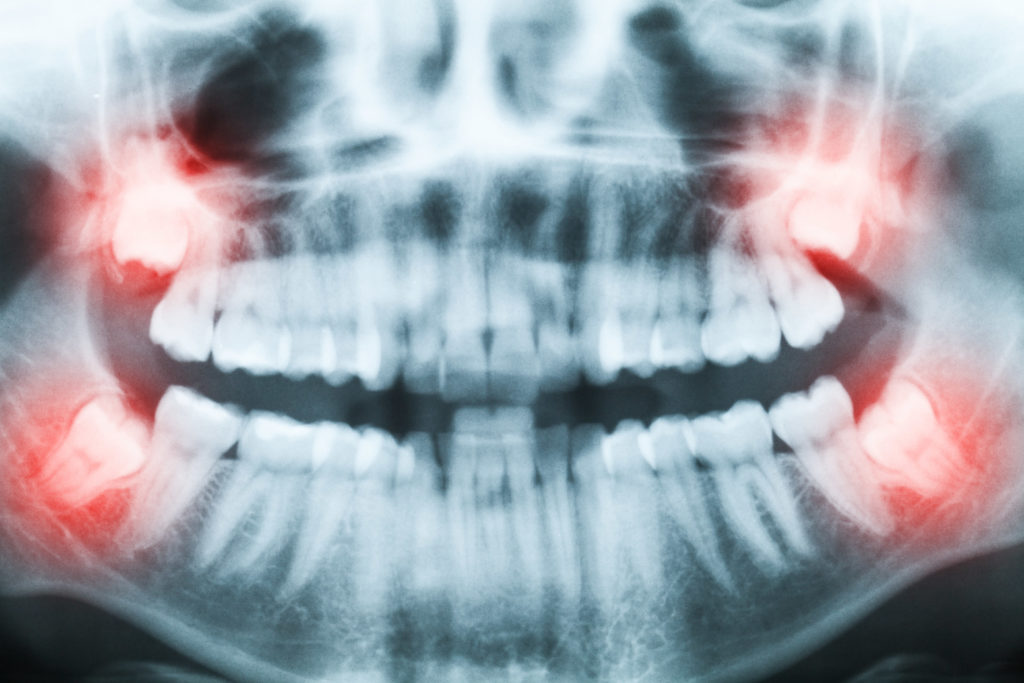
- Panoramic X-Rays: Panoramic x-rays are unique in the way that they show your entire mouth, both top and bottom teeth all in one image. Instead of often being done in a chair like the bitewing x-ray, the panoramic x-ray requires the patient to stand up inside the machine. The x-ray then rotates around you to create one continuous image of your mouth. Panoramic images are used for several purposes but most likely they’ll be used for orthodontic planning involving braces or as a part of the wisdom teeth removal process.
- Periapical X-Rays: Periapical x-rays are similar to bitewing x-rays in the sense that they only look at a few teeth at a time. However periapical x-rays are a little more intricate because they show the entire tooth, from crown to root. These types of x-rays are crucial for analyzing and diagnosing dental abscesses.
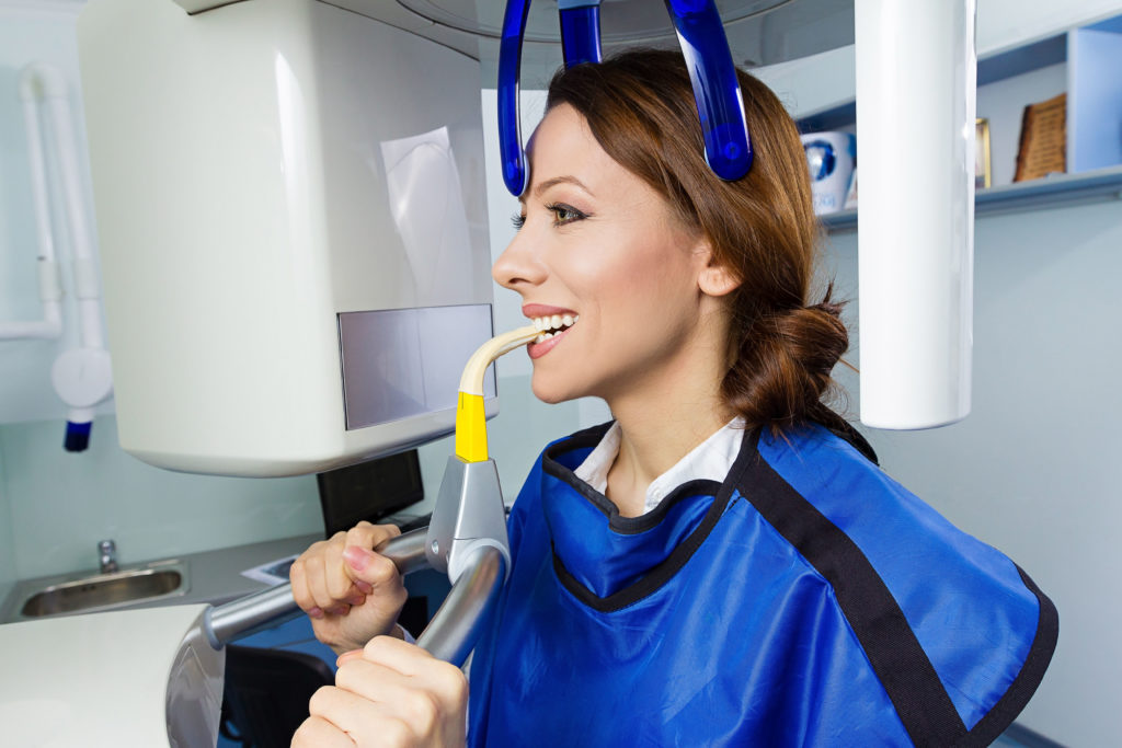
- Cone Beam CT: The “CT” in Cone Beam CT stands for “computed tomography” and is essentially a computer assisted 3-D x-ray model. Similar to the Panoramic X-Ray, the patient will typically stand within the machine as it rotates around them. The images are then sent to a computer where a special program helps convert the images into a 3-D model version. Cone Beams are typically the least common type of x-ray and are rarely used on patients. More then not cone beams are for the planning and configuration of dental implants.
X-Ray Safety
It’s widely believed that x-rays are unsafe to use because they expose you to excessive levels of harmful radiation. While there is truth in the fact that x-rays expose you to radiation, the levels of radiation are so miniscule that they are practically harmless.
However, the claim that x-rays are dangerous isn’t to be completely ignored. Exposure to any radiation no matter how big or small should be closely examined. Dentists will limit your exposure as much as possible and will not take your x-ray unless it serves a purpose. Even stricter precautions are made when addressing pregnant women.
Although they weren’t always this effective, the good news is that nowadays x-rays are extremely safe to use. Over the years many alterations have been made to the overall design of the x-ray to make it safer. Some of which include but aren’t limited to:
- Decreasing The Size of The X-Ray Window: This basically means x-rays have been polished to only interact with very small, very specific areas of your mouth to reduce the amount of radiation you are exposed to.
- Faster Film Processes: Modernized film is much quicker than traditional films meaning a patient doesn’t have to be exposed nearly as long for an image to be made.
- Digital Radiography: Often regarded as the most significant advancement for x-ray technology, computers assist x-rays post process and can sort of “fill in the gaps” so that less has to be done in the actual exposure process. Digital radiography also helps professionals develop 3-D models and more accurate, effective x-ray images.
- Safety Regulations: Certain standards and laws have been made and improved over the years to ensure that x-ray usage is safer than ever before.
- Lead shields: Aprons lined with lead are optimized to even further protect patients from radiation. Lead has very unique properties of it that help it to deter radiation and create for a much safer x-ray experience.
As you can see, x-ray technology has come quite a long way since 1895. With all the precautions and advancements made over the past century, there is no reason anyone should feel at risk when going to the dentist. Our x-rays are not just extremely safe but they are extremely accurate as well and they ensure that our professionals here at Mill Dam Dentistry can offer you the best services possible. For more information on dental topics such as these check out our other informative dental blogs.